The radial nerve is the terminal continuation of the posterior cord of the brachial plexus. It contains fibers from nerve roots C5 – T1. From the axilla region, the radial nerve travels down the arm and innervates the triceps muscle. The radial nerve then descends down the arm, travelling in a shallow depression within the surface of the humerus, known as the radial groove.
The radial nerve wraps around the humerus laterally and enters the forearm anterior to the lateral epicondyle of the humerus, through the cubital fossa. The nerve then terminates by dividing into two branches:
- Deep branch (motor) – innervates the muscles in the posterior compartment of the forearm.
- Superficial branch (sensory) – contributes to the cutaneous innervation of the dorsal hand and fingers.
The loss of radial nerve function typically has a significant negative motor impact on an individual.
Neuropraxia
Neuropraxia is a temporary interruption of conduction without loss of axonal continuity.
In neurapraxia, there is a physiologic block of nerve conduction in the affected axons.
Other characteristics:
- It is the mildest type of peripheral nerve injury.
- There are sensory-motor problems distal to the site of injury.
- Everything is still intact.
Axonotmesis
Axonotmesis involves loss of the relative continuity of the axon and its covering of myelin, but preservation of the connective tissue framework of the nerve (the encapsulating tissue, the epineurium and perineurium, are preserved).
- There are sensory and motor deficits distal to the site of lesion.
Neurotmesis
Neurotmesis is a total severance or disruption of the entire nerve fiber. A peripheral nerve fiber contains an axon (Or long dendrite), myelin sheath (if existence), their Schwann cells, and the endoneurium. Neurotmesis may be partial or complete.
- Sensory-motor problems and autonomic function defects are severe.
The radial nerve can be damaged in the axilla region by a dislocation at the shoulder joint, or a fracture of the proximal humerus. Occasionally, it is injured via excessive pressure on the nerve within the axilla (use of crutches) or falling asleep with the arm draped over a sofa.
- Motor functions – the triceps brachii and muscles in posterior compartment are affected. The patient is unable to extend at the elbow, wrist, and fingers. Unopposed flexion of wrist occurs, known as wrist-drop.
- Sensory functions – all four cutaneous branches of the radial nerve are affected. There will be a loss of sensation over the lateral and posterior arm, posterior forearm, and dorsal surface of the lateral three and a half
The radial nerve can be damaged in the mid -shaft of the humerus, commonly during a fall and subsequent humeral fracture.
- Motor functions – The radial nerve is most likely to be damaged in humerus fractures that have a lateral displacement of the distal fracture segment, as the nerve is tethered to the bone and cannot withstand the forces applied to it as a result of the displacement. On physical exam, patients with a radial nerve injury may have wrist drop (loss or weakness of wrist extensors), loss or weakness of finger extension.
- Up to 90% of patients with a closed humeral fracture with radial nerve injury will have a resolution of neuropraxia within three to four months following the injury.
- Sensory decreased or absent sensation to the posterior forearm, digits 1 to 3, and the radial half of the fourth digit.
Orthoses
There are many different styles and designs of orthoses for radial nerve palsy. The goals are always the same:
- Provide wrist support or assisted wrist extension
- Provide assisted Metacarpal-phalangeal (MCP) joint extension while allowing full finger flexion.
Low-Profile Design
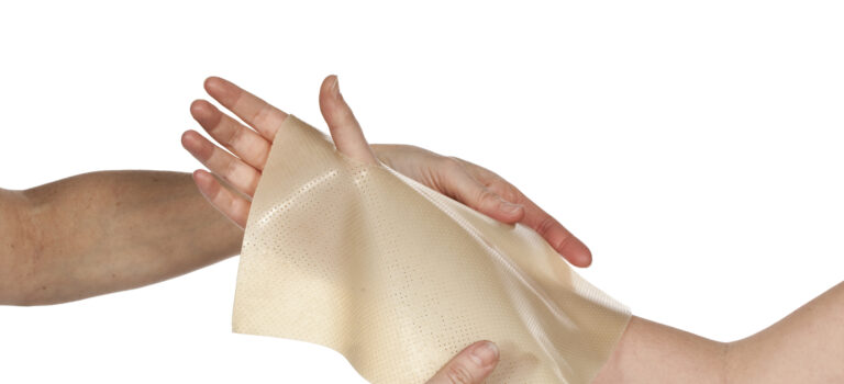
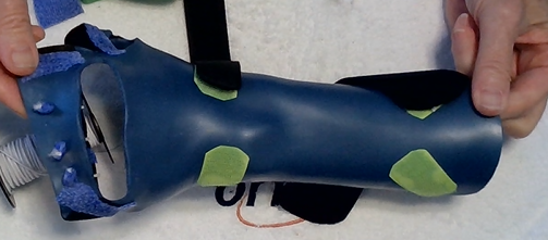
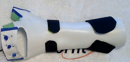
We have created a low-profile radial nerve palsy orthosis! The beauty of this design is that it does not include high outriggers but maintains the MCP joints in extension with waistband elastic. It allows for full finger flexion and can accommodate the MCP joints in flexion with an opening. The wrist is maintained in extension, but a simple modification allows for assisted wrist extension with adjustable tension.
Source: Peck, J., & Ollason, J. (2015). Low profile radial nerve palsy orthosis with radial and ulnar deviation. Journal of Hand Therapy, 28(4), 421-424.
Sign up here to get a link to the video demonstrations and learn how to fabricate this low-profile design.


Written by Debby Schwartz, OTD, OTR/L, CHT
Physical Rehabilitation Product and Educational Specialist at Orfit Industries America.
Debby is a certified hand therapist with over 36 years of clinical experience. She completed her Doctorate of Occupational Therapy at Rocky Mountain University of Health Professions in 2010. She has worked at Orfit Industries America as Product and Educational Specialist since 2007.
Debby is also an adjunct professor at the Occupational Therapy Department of Touro University, School of Health Sciences, and at the Occupational Therapy Department at Yeshiva University, Katz School of Science and Health in NYC. She has written many book chapters in the field of hand therapy and multiple articles for hand therapy journals, including the ASHT Times and the Journal of Hand Therapy. She has published a new textbook on orthotic fabrication together with Dr. Katherine Schofield, entitled “Orthotic Design and Fabrication for the Upper Extremity: A Practical Guide”.
Debby’s Google Scholar